Australian researchers have made a surprise discovery that could rewrite our understanding of the role programmed cell death plays in embryonic development and congenital birth defects.
The team showed that, while programmed cell death – or apoptosis – is essential for healthy development overall, many organs and tissues do not require apoptosis to develop normally.
The study, published today in the high-ranking journal Cell, also suggested that abnormalities in cell death processes are likely to contribute to some common birth defects in humans, such as spina bifida, heart vessel defects and cleft palate.
Walter and Eliza Hall Institute researchers Dr Francine Ke, Dr Hannah Vanyai, Dr Angus Cowan, Associate Professor Anne Voss and Professor Andreas Strasser led the research.

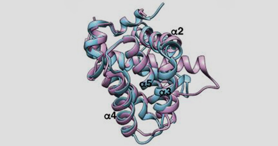
Experiments to determine the crystal structure of a key 'pro-death' protein, BOK, were carried out using the MX beamline at the Australian Synchrotron.
Cell death link to birth defects
Programmed cell death, also known as apoptosis, is a normal process that rids the body of sick, damaged or unwanted cells in a controlled way, limiting side effects and damage to the body.
Apoptosis was first described as having a role in embryonic development in the 1940s. Over the past 70 years, numerous studies have implicated apoptosis as playing a crucial role in most stages and tissues during development.
However, in this new study, it became clear that apoptosis was not as critical during development as previously thought, Dr Ke said.
“Rather, apoptosis was essential at specific places and times during development, but unnecessary in others. We identified the tissues and organs that critically require apoptosis to develop normally, and made the surprise discovery that many do not require it at all,” Dr Ke said.
The finding provides clear clues about a link between abnormalities in programmed cell death and some common congenital birth defects, including spina bifida, heart vessel defects and cleft palate.
“Our research showed that when cell death is not functioning properly, it commonly leads to defects in neural tube development, for example spina bifida, heart vessel defects and facial abnormalities, such as cleft palate,” Dr Ke said.
Surprise discovery
Associate Professor Voss said she thought the biggest surprise came from the discovery of the tissues that did not require apoptosis at all for normal development.
Dr Cowan said the study confirmed that BOK acted as a pro-death protein. “In this paper we have solved the structure of BOK using the Australian Synchrotron, and once and for all confirmed that BOK is a pro-death protein that plays an important role in apoptosis,” he said.
“For some time, it has been a widely-held belief that programmed cell death is necessary for the shaping of certain tissues and structures during development. But, to our surprise, many tissues in which programmed cell death was – for very good reasons – considered absolutely essential, it is not required at all," Associate Professor Voss said.
“For example, apoptosis was thought to play a particularly important role in ‘hollowing out’ of ducts in internal organs during development.
"However we have shown that – in the absence of apoptosis – most tissues and organs develop normally. I think it may surprise researchers to learn just how precise and limited the effects of apoptosis are in embryonic development."
The research was supported by the Australian National Health and Medical Research Council, Cancer Council Victoria, Lady Tata Memorial Trust, Leukaemia Foundation of Australia, Leukemia & Lymphoma Society (US) and the Victorian Government.
